Introduction
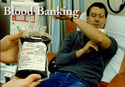
You can make a deposit or withdrawal from this bank. The best news...no ATM charges! Transfusion medicine deals with the use of blood products in a therapeutic setting. Since the very first blood product transfusion, there has been concern over the safety of the blood supply. It is a perishable product with stringent guidelines that must be followed by every blood bank and laboratory that deals with blood products. The following table highlights the current risk associated with the transfusion of a single unit of blood, platelets, or cryoprecipitate. To give an accurate comparison of the relative risks, the probability of a fatal car accident is approximately 1:6000 for each year we drive to work in Los Angeles.
VIRUS TESTED CURRENT RISK PREVIOUS RISK HIV 1:2,135,000 1:500,000 HEPATITIS C 1:1,935,000 1:100,000 HEPATITIS B 1:205,000 1:63,000 HTLV-I/II 1:2,993,000 1:500,000 Adopted from Transfusion 2002;42:975-979.
OUTLINE
Autologous Blood Products Fresh Frozen Plasma Leukocyte Reduced Transfusions Platelets Red Blood Cells Clinical Utility Transfusion Reactions Commonly Used Terms Internet Links
BLOOD PRODUCTS CHARACTERIZATION AUTOLOGOUS DONATION
- Storage and transfusion of infected autologous blood or components: a survey of North American laboratories.
Shulman IA, Osby M.
Department of Pathology, Keck School of Medicine, University of Southern California, Los Angeles County-USC Medical Center, Los Angeles, California, USA.
Arch Pathol Lab Med. 2005 Aug;129(8):981-3. Abstract quote
CONTEXT: Many patients request that autologous blood or components be collected and available for use during scheduled surgical or invasive medical procedures to avoid exposure to human immunodeficiency virus (HIV), hepatitis B virus (HBV), and hepatitis C virus (HCV) from allogeneic transfusions. Some patients from whom autologous blood is collected are themselves infected with HIV, HBV, or HCV. However, unlike HIV-, HBV-, or HCV-infected allogeneic blood and components, which must be excluded from the community blood supply, infected autologous blood and components are allowed to be stored in hospitals and transfused back to the patients (autologous donors) from whom the blood was collected. Although the transfusion of HIV-, HBV-, or HCV-infected autologous blood or components does not present a risk to the autologous donor, such a transfusion presents a risk to other patients, considering that at least 1 in every 25,000 transfusions are administered to the wrong individual.
OBJECTIVE: To determine if hospital transfusion services store and/or transfuse autologous blood or components infected with HIV, HBV, and/or HCV.
DESIGN: An educational enhancement subsection of a College of American Pathologists Proficiency Testing Survey (J-C 2003) assessed transfusion service practices for storing and/or transfusing HIV-, HBV-, and HCV-infected autologous blood and components.
SETTING AND PARTICIPANTS: A total of 4251 participants were asked whether they stored and/or transfused autologous blood or components and whether these stored blood products included those that were infected with HIV, HBV, or HCV. RESULTS: Of the 4251 survey respondents, 3561 provided data regarding their autologous blood and component storage and/or transfusion practices. A total of 2988 participants reported that they store and/or transfuse autologous blood or components. A total of 2390 respondents reported that they do not test autologous donations collected in their own institution for evidence of infection with HIV, HBV, or HCV. Most survey participants reported that even if an autologous donation is tested and found to be infected they would still be willing to store and transfuse the blood component, according to which agent was causing the infection: HIV (n = 1867), HBV (n = 2158), or HCV (n = 2233).
CONCLUSION: Most North American hospitals do not test autologous blood donations that they collect in their own institution for evidence of infection with HIV, HBV, or HCV, leading to the conclusion that infected autologous blood components are being stored and transfused. Even when autologous donations are tested and found to be infected with HIV, HBV, or HCV, most North American hospitals would be willing to store and/or transfuse the infected autologous blood components.FRESH FROZEN PLASMA
Quality indicators of fresh frozen plasma and platelet utilization.Novis DA, Renner S, Friedberg RC, Walsh MK, Saladino AJ.
Department of Pathology, Wentworth-Douglass Hospital, Dover, NH 03820, USA.
Arch Pathol Lab Med 2002 May;126(5):527-32 Abstract quote OBJECTIVE: To determine the normative rates of expiration and wastage for units of fresh frozen plasma (FFP) and platelets (PLTs) in hospital communities throughout the United States, and to examine hospital blood bank practices associated with more desirable (lower) rates.
DESIGN: In 3 separate studies, participants in the College of American Pathologists Q-Probes laboratory quality improvement program collected data retrospectively on the numbers of units of FFP and PLTs that expired (outdated) prior to being used and that were wasted due to mishandling. Participants also completed questionnaires describing their hospitals' and blood banks' laboratory and transfusion practices.
SETTING AND PARTICIPANTS: One thousand six hundred thirty-nine public and private institutions, more than 80% of which were known to be located in the United States.
MAIN OUTCOME MEASURES: Quality indicators of FFP and PLT utilization: the rates of expiration and wastage of units of FFP and PLTs.
RESULTS: Participants submitted data on 8 981 796 units of FFP and PLTs. In all 3 studies, aggregate combined FFP and PLT expiration rates ranged from 5.8% to 6.4% and aggregate combined FFP and PLT wastage rates ranged from 2.0% to 2.5%. Among the top-performing 10% of participants (90th percentile and above), FFP and PLT expiration rates were 0.6% or lower and FFP and PLT wastage rates were 0.5% or lower. Among the bottom-performing 10% of participants (10th percentile and below), expiration rates were 13.8% or higher and wastage rates were 6.8% or higher. We were unable to associate selected hospital characteristics or blood bank practices with lower rates of FFP and PLT utilization.
CONCLUSIONS: The rates of FFP and PLT expiration and wastage vary greatly among hospitals in the United States. Hospital blood bank personnel are capable of achieving FFP and PLT expiration and wastage rates below 1%.
LEUKOCYTE REDUCED TRANSFUSIONS
Clinical outcomes following institution of universal leukoreduction of blood transfusions for premature infants.Fergusson D, Hebert PC, Lee SK, Walker CR, Barrington KJ, Joseph L, Blajchman MA, Shapiro S.
Departments of Epidemiology and Biostatistics and Pediatrics, McGill University, Montreal, Quebec.
JAMA 2003 Apr 16;289(15):1950-6 Abstract quote CONTEXT: Leukocytes present in stored blood products can have a variety of biological effects, including depression of immune function, thereby increasing nosocomial infections and possibly resulting in organ failure and death. Premature infants, given their immature immune state, may be uniquely predisposed to the effects of transfused leukocytes.
OBJECTIVE: To evaluate the clinical outcomes following implementation of a universal prestorage red blood cell (RBC) leukoreduction program in premature infants admitted to neonatal intensive care units (NICUs).
DESIGN AND SETTING: Retrospective before-and-after study conducted in 3 Canadian tertiary care NICUs from January 1998 to December 2000.
PATIENTS: A total of 515 premature infants weighing less than 1250 g who were admitted to the NICU, received at least 1 RBC transfusion, and survived at least 48 hours were enrolled. The intervention group consisted of infants admitted in the 18-month period following the introduction of universal leukoreduction (n = 247) and the control group consisted of infants admitted during the 18 months prior to the introduction of universal leukoreduction (n = 268).
MAIN OUTCOME MEASURES: Primary outcomes were nosocomial bacteremia and NICU mortality, compared before and after implementation of universal leukoreduction using multivariate regression. Secondary outcomes included bronchopulmonary dysplasia, retinopathy of prematurity, necrotizing enterocolitis, and intraventricular hemorrhage.
RESULTS: The proportion of infants who acquired bacteremia after an RBC transfusion was 79/267 (29.6%) in the nonleukoreduction period and 63/246 (25.6%) in the leukoreduction period. For NICU mortality, there were 45 deaths (16.8%) in the nonleukoreduction period and 44 deaths (17.8%) in the leukoreduction period. The adjusted odds ratio (OR) for bacteremia was 0.59 (95% confidence interval [CI], 0.34-1.01) and for mortality was 1.22 (95% CI, 0.59-2.50). The adjusted ORs for bronchopulmonary dysplasia and retinopathy of prematurity were 0.42 (95% CI, 0.25-0.70) and 0.56 (95% CI, 0.33-0.93), respectively. The adjusted ORs for necrotizing enterocolitis and grade 3 or 4 intraventricular hemorrhage were 0.39 (95% CI, 0.17-0.90) and 0.65 (95% CI, 0.35-1.19), respectively. The adjusted OR for a composite measure of any major neonatal morbidity was 0.31 (95% CI, 0.17-0.56). Crude and adjusted rates for all secondary outcomes suggest that leukoreduction was associated with improved outcomes.
CONCLUSION: Implementation of universal prestorage leukoreduction was not associated with significant reductions in NICU mortality or bacteremia but was associated with improvement in several clinical outcomes in premature infants requiring RBC transfusions.
Clinical outcomes following institution of the canadian universal leukoreduction program for red blood cell transfusions.Hebert PC, Fergusson D, Blajchman MA, Wells GA, Kmetic A, Coyle D, Heddle N, Germain M, Goldman M, Toye B, Schweitzer I, VanWalraven C, Devine D, Sher GD.
University of Ottawa Centre for Transfusion Research, and Clinical Epidemiology Program of the Ottawa Health Research Institute, Ottawa, Ontario.
JAMA 2003 Apr 16;289(15):1941-9 Abstract quote CONTEXT: A number of countries have implemented a policy of universal leukoreduction of their blood supply, but the potential role of leukoreduction in decreasing postoperative mortality and infection is unclear.
OBJECTIVE: To evaluate clinical outcomes following adoption of a national universal prestorage leukoreduction program for blood transfusions.
DESIGN, SETTING, AND POPULATION: Retrospective before-and-after cohort study conducted from August 1998 to August 2000 in 23 academic and community hospitals throughout Canada, enrolling 14 786 patients who received red blood cell transfusions following cardiac surgery or repair of hip fracture, or who required intensive care following a surgical intervention or multiple trauma.
INTERVENTION: Universal prestorage leukoreduction program introduced by 2 Canadian blood agencies. A total of 6982 patients were enrolled during the control period and 7804 patients were enrolled following prestorage leukoreduction. MAIN
OUTCOME MEASURES: All-cause in-hospital mortality and serious nosocomial infections (pneumonia, bacteremia, septic shock, all surgical site infections) occurring after first transfusion and at least 2 days after index procedure or intensive care unit admission. Secondary outcomes included rates of posttransfusion fever and antibiotic use.
RESULTS: Unadjusted in-hospital mortality rates were significantly lower following the introduction of leukoreduction compared with the control period (6.19% vs 7.03%, respectively; P =.04). Compared with the control period, the adjusted odds of death following leukoreduction were reduced (odds ratio [OR], 0.87; 95% confidence interval [CI], 0.75-0.99), but serious nosocomial infections did not decrease (adjusted OR, 0.97; 95% CI, 0.87-1.09). The frequency of posttransfusion fevers decreased significantly following leukoreduction (adjusted OR, 0.86; 95% CI, 0.79-0.94), as did antibiotic use (adjusted OR, 0.90; 95% CI, 0.82-0.99).
CONCLUSION: A national universal leukoreduction program is potentially associated with decreased mortality as well as decreased fever episodes and antibiotic use after red blood cell transfusion in high-risk patients.
Leukocyte-reduced transfusions in cardiac surgery results of an implementation trial.Blumberg N, Heal JM, Cowles JW, Hicks GL Jr, Risher WH, Samuel PK, Kirkley SA.
Department of Pathology & Laboratory Medicine, University of Rochester Medical Center, NY 14642, USA.
Am J Clin Pathol 2002 Sep;118(3):376-81 Abstract quote An implementation trial of leukocyte-reduced transfusions in cardiac surgery (primary coronary artery bypass graft and valve replacement) was performed from July to December 1998; comparisons were made with data from the same period in 1997.
Patients from both periods were similar in important preoperative and intraoperative variables (age, sex, weight, number of units of RBCs transfused, ejection fraction). The mean total number of complications was statistically significantly decreasedfrom 0.26 complications per patient in the non-leukocyte-reduced to 0.19 in the leukocyte-reduced recipients. Overall, the mean +/- ISD costs of care per patient decreasedfrom 1997 ($27,615 +/- $33,973) to 1998 ($27,038 +/- $24,107).
Mean costs decreased $1,700 per patient for recipients of leukocyte-reduced blood in 1998 compared with recipients of non-leukocyte-reduced blood in 1997 Mean costs increased $4,000 per patient in patients who did not receive transfusions in 1998 compared with 1997. Hospitalization costs decreased when leukocyte-reduced transfusions were implemented for patients undergoing cardiac surgery in our institution. Implementation of leukocyte reduction may be cost neutral or cost saving in at least some settings.
PLATELETS
Bacterial contamination of platelet units: a case report and literature survey with review of upcoming american association of blood banks requirements.
Burns KH, Werch JB.
Department of Pathology, Baylor College of Medicine, Houston, Tex, USA.
Arch Pathol Lab Med. 2004 Mar;128(3):279-81. Abstract quote
The most common transfusion-associated infectious risk in the United States today is bacterial contamination of platelet components. Bacterial contamination is estimated to occur at an incidence of 1:1000 to 1:3000 in platelet units, with severe episodes estimated to occur in about one sixth of contaminated products. Increased awareness and prompt reaction of the medical team can greatly affect the outcome and save a patient's life.
The following case history illustrates this issue. A young woman developed chills and rigors while receiving 1 unit of leuko-reduced apheresis platelets for severe thrombocytopenia. The transfusion was stopped, blood cultures were drawn, and the patient developed clinical signs of sepsis. Cultures of both the platelet unit and the patient's blood revealed coagulase-negative Staphylococcus. Microbial susceptibilities in both samples were identical. Pretransfusion blood cultures taken from the patient earlier that day were negative. The platelet unit had been stored for 5 days.
We review this case and the literature describing the persistent problem of platelet unit contamination and at the same time highlight the efforts now directed by the American Association of Blood Banks and College of American Pathologists to address this issue. Although there is no uniform approach to dealing with bacterial contamination of platelets, the American Association of Blood Banks and the College of American Pathologists have promulgated new accreditation requirements in an effort to prevent bacterial sepsis associated with platelet transfusion. A new American Association of Blood Banks standard, which will be effective March 1, 2004, requires a combination of strategies both to limit the initial inoculation of bacteria into the blood component and to detect subsequent growth at room temperature (American Association of Blood Banks Association Bulletin #03-12).
The new College of American Pathologists Checklist question, which became effective in December 2003, is a Phase 1 requirement that calls for inspected facilities to have a platelet bacteria detection method in place.RED BLOOD CELLS
Quality indicators of blood utilization: three College of American Pathologists Q-Probes studies of 12,288,404 red blood cell units in 1639 hospitals.Novis DA, Renner S, Friedberg R, Walsh MK, Saladino AJ.
Department of Pathology, Wentworth-Douglass Hospital, Dover, NH 03820, USA.
Arch Pathol Lab Med 2002 Feb;126(2):150-6 Abstract quote OBJECTIVES: To determine the normative rates of blood unit crossmatched to transfused (C:T) ratios, red blood cell (RBC) unit wastage, and RBC unit expiration that exist in hospital communities throughout the United States, and to examine hospital blood bank practices associated with more desirable (lower) rates.
DESIGN: In 3 separate studies, participants in the College of American Pathologists Q-Probes laboratory quality improvement program collected data retrospectively on the number of transfusion crossmatches performed in their institutions and the number of RBC-containing units that were transfused into patients, the number of units that expired (outdated) prior to being utilized, and the number that were wasted due to mishandling. Participants also completed questionnaires describing their hospitals' and blood banks' laboratory and transfusion practices.
SETTING AND PARTICIPANTS: One thousand six hundred thirty-nine public and private institutions, well more than 80% of which were known to be located in the United States.
MAIN OUTCOME MEASURES: Quality indicators of blood utilization (namely, the C:T ratio, the rate of RBC unit expiration, and the rate of RBC unit wastage).
RESULTS: Participants submitted data on 12,288,404 RBC unit transfusions. The C:T ratios were 1.5 or less in the top-performing 10% of participating institutions (90th percentile and above), 1.8 to 1.9 in the midrange of participating institutions (50th percentile), and 2.4 or greater in the bottom-performing 10% of participating institutions (10th percentile and below). Red blood cell unit expiration rates were 0.1% or less at the 90th percentile and above, 0.3% to 0.9% at the 50th percentile, and 3.5% or greater at the 10th percentile and below. Red blood cell unit wastage rates were 0.1% or less at the 90th percentile and above, 0.1% to 0.4% at the 50th percentile, and 0.7% or greater at the 10th percentile and below. Depending on which quality indicator was examined, lower values (ie, better performances) were found in institutions that had fewer than 200 hospital beds, no teaching programs, no on-site full-time medical directors of transfusion services, did not utilize maximum surgical blood order schedules, set C:T threshold goals of 2.0 or less, monitored categories of health care workers responsible for RBC wastage, monitored requests for RBC components by transfusion indication, did not accept short-dated units from blood distribution centers, and if they did accept short-dated units, were allowed to return those units to the distribution centers.
CONCLUSIONS: Hospital blood bank personnel can achieve C:T ratios below 2.0, RBC unit expiration rates below 1.0%, and RBC unit wastage rates below 0.5%. Lower C:T ratios and/or RBC unit expiration rates were associated with blood bank personnel setting C:T thresholds of 2.0 or less, monitoring requests for blood components by transfusion indication criteria, monitoring categories of health care workers responsible for blood wastage, not accepting short-dated units from blood distribution centers, and if short-dated units were accepted, being allowed to return those units to the blood distribution center. These practices were not associated with lower blood wastage rates.
CHARACTERIZATION
GENERAL
ALLERGIC
Allergic transfusion reactions: an evaluation of 273 consecutive reactions.Domen RE, Hoeltge GA.
Cleveland Clinic, Department of Clinical Pathology, Section of Transfusion Medicine, Cleveland, Ohio, USA.
Arch Pathol Lab Med 2003 Mar;127(3):316-20 Abstract quote
CONTEXT: Allergic reaction to transfusion is common. However, the review of a large series of allergic transfusion reactions has not been performed.
OBJECTIVE: To review a large series of allergic transfusion reactions.
DESIGN: A retrospective review of all reported and evaluated transfusion reactions during a 9-year period at 1 institution was performed. Associated clinical signs and symptoms were evaluated.
SETTING: Large, tertiary-care teaching hospital.
RESULTS: A total of 1613 adverse reactions to transfusion were evaluated. Allergic transfusion reactions accounted for 17% (273 of 1613) of the transfusion reactions. Severe allergic reactions (anaphylaxis, anaphylactoid signs and symptoms, and/or hypotension) were observed in 21 patients (7.7% of allergic reactions, or 1.3% of all transfusion reactions). Serum tryptase, a marker for anaphylaxis, was measured in 1 patient and determined to be borderline elevated. Five patients experienced allergic transfusion reactions to autologous red cell transfusions. One patient experienced hives during the transfusion of a major ABO mismatched red blood cell. A wide variety of skin manifestations were observed, but 26 (9.5%) patients did not have skin manifestations. Allergic transfusion reactions were estimated to occur in approximately 1 in 4124 blood components transfused, or 1 in 2338 transfusion episodes. Severe allergic reactions occurred in approximately 1 in 30,281 transfusions. No deaths directly attributable to transfusion were observed in this patient group.
CONCLUSIONS: The clinical presentation of allergic transfusion reactions was quite variable, and the pathophysiology remains unclear. Recommendations for clinical evaluation and therapy remain problematic and often empirical.
FEBRILE NONHEMOLYTIC TRANSFUSION REACTION
- Febrile nonhemolytic transfusion reactions. Management by premedication and cost implications in adult patients.
Ezidiegwu CN, Lauenstein KJ, Rosales LG, Kelly KC, Henry JB.
Department of Pathology, State University of New York Upstate Medical University, Syracuse, NY 13210, USA.
Arch Pathol Lab Med. 2004 Sep;128(9):991-5. Abstract quote
CONTEXT: Febrile nonhemolytic transfusion reactions (FNHTRs) cause unwelcome interruptions during the course of blood product transfusions and necessitate measures to verify the nature of the reaction and to exclude certain dangerous reactions, such as hemolytic and septic phenomena.
OBJECTIVE: To examine transfusion medicine data to determine the clinical implications of the routine administration of antipyretic medication to adult patients before transfusion for the prevention of FNHTRs.
DESIGN: A retrospective review was conducted of FNHTR data during 5 years (1998-2002), and a determination was made of the cost of a transfusion complicated by an FNHTR. In addition, a comparative cost analysis was performed using our data and published data on the incidence of FNHTRs. The clinical implications of medication with respect to possible drug-induced adverse effects were assessed, as well as the potential interference with diagnosing other forms of transfusion reactions and the mitigation of the clinical effect of an FNHTR.
RESULTS: For nearly 120,000 U of transfused blood components, approximately 80% of which were preceded by antipyretic medication during the study period, the overall incidence of FNHTR was found to be 0.09%. Furthermore, there was no evidence of antipyretic-associated complications, nor any evidence that antipyretics prevented the recognition of other more dangerous complications of transfusions.
CONCLUSION: Our findings indicate that this practice provides significant advantages to the recipient of a transfusion, but does not appear to yield significant cost benefits for the health care provider.WARM REACTIVE AUTOANTIBODIES
- Warm reactive autoantibodies: clinical and serologic correlations.
Wheeler CA, Calhoun L, Blackall DP.
Department of Pathology and Laboratory Medicine, University of California, Los Angeles.
Am J Clin Pathol. 2004 Nov;122(5):680-5. Abstract quote
Warm reactive autoantibodies are encountered relatively frequently in tertiary care hospitals.
We studied 100 consecutive patients with warm autoantibodies to correlate their clinical and serologic features. Study patients (56 male, 44 female) had various diagnoses and a mean age of 53.5 years (range, 3-90 years). Autoimmune hemolysis was documented in 29 patients; 20 patients (69%) in this subset had diseases classically associated with warm autoimmune hemolytic anemia (hematologic and autoimmune disorders). All study patients demonstrated IgG on their RBCs (direct antiglobulin test [DAT] reactivity range, microscopic to 4+); 49 also demonstrated C3 (reactivity range, microscopic to 3+). The DAT for IgG was 2+ or more in 25 (86%) of 29 patients with hemolysis; the DAT for IgG was 1+ or less in 45 (63%) of 71 patients without hemolysis. In patients with hemolysis, 21 (72%) of 29 had a DAT reactive for C3.
These findings may be useful in determining the clinical significance of warm autoantibodies and the extent to which patients should be followed up for hemolysis.
Henry JB. Clinical Diagnosis and Management by Laboratory Methods. Twentieth Edition. WB Saunders. 2001.
Rosai J. Ackerman's Surgical Pathology. Ninth Edition. Mosby 2004.
Sternberg S. Diagnostic Surgical Pathology. Fourth Edition. Lipincott Williams and Wilkins 2004.
Robbins Pathologic Basis of Disease. Seventh Edition. WB Saunders 2005.
DeMay RM. The Art and Science of Cytopathology. Volume 1 and 2. ASCP Press. 1996.
Weedon D. Weedon's Skin Pathology Second Edition. Churchill Livingstone. 2002
Fitzpatrick's Dermatology in General Medicine. 5th Edition. McGraw-Hill. 1999.
Weiss SW and Goldblum JR. Enzinger and Weiss's Soft Tissue Tumors. Fourth Edition. Mosby 2001.
Transfusion Related Acute Lung Injury
Basic Principles of Disease
Learn the basic disease classifications of cancers, infections, and inflammation
Commonly Used Terms
This is a glossary of terms often found in a pathology report.Diagnostic Process
Learn how a pathologist makes a diagnosis using a microscopeSurgical Pathology Report
Examine an actual biopsy report to understand what each section meansSpecial Stains
Understand the tools the pathologist utilizes to aid in the diagnosisHow Accurate is My Report?
Pathologists actively oversee every area of the laboratory to ensure your report is accurateGot Path?
Recent teaching cases and lectures presented in conferences
Last Updated August 4, 2005
Send mail to The Doctor's Doctor with questions or comments about this web site.
Read the Medical Disclaimer.
Copyright © The Doctor's Doctor