
Background
This rare tumor occurs predominantly in an intraoral location, with the palate the most common site. It is a tumor that may mimic many benign and malignant salivary gland tumors and for many years, was probably misdiagnosed as tumors such as adenoid cystic carcinoma and mixed tumors.
OUTLINE
EPIDEMIOLOGY CHARACTERIZATION SYNONYMS Lobular carcinoma of the minor salivary glands
Low grade papillary carcinoma of the palate
Polymorphous low-grade adenocarcinoma of minor salivary glands
Terminal duct carcinoma of the minor salivary glandsINCIDENCE Rare AGE RANGE-MEDIAN 22-71 years
Median 54 years
- Polymorphous low-grade adenocarcinoma--a rare and aggressive entity in adolescence.
Kumar M, Stivaros N, Barrett AW, Thomas GJ, Bounds G, Newman L.
Maxillofacial Unit, University College Hospital, London WC1E 6AU, UK.
Br J Oral Maxillofac Surg. 2004 Jun;42(3):195-9. Abstract quote
Polymorphous low-grade adenocarcinoma (PLGA) is an uncommon tumour that usually affects the minor salivary glands, particularly in the palate.
It is rare in young patients, and here we report a case in a teenage girl. She presented at the age of 16, although the lesion had been noticed 2 years previously. The tumour showed histopathological features of PLGA, but recurred locally, behaved aggressively, and ultimately metastasised to cervical lymph nodes.
This was accompanied by an altered histological picture, with a papillary cystic pattern and necrosis becoming progressively more prominent.
PLGA is not always a low-grade lesion and some tumours, notably those with a papillary cystic growth pattern, may require more aggressive treatment.
Slight female predominance
PATHOGENESIS CHARACTERIZATION CHROMOSOMAL ABNORMALITIES
- Polymorphous low grade adenocarcinoma with distant metastases and deletions on chromosome 6q23-qter and 11q23-qter: a case report.
Hannen EJ, Bulten J, Festen J, Wienk SM, de Wilde PC.
Department of Oral and Maxillofacial Surgery, University Hospital Nijmegen, PO Box 9101, 6500 HB Nijmegen, The Netherlands.
J Clin Pathol. 2000 Dec;53(12):942-5. Abstract quote
Polymorphous low grade adenocarcinomas (PLGAs) are thought to be indolent tumours that are localised preferentially to the palate and affect the minor salivary glands almost exclusively. Metastases to locoregional lymph nodes occur in only 6-10% of cases. Recently, two cases of PLGA with microscopically confirmed distant metastases have been reported.
This study reports a third case of PLGA with histologically and immunohistochemically confirmed distant metastases. It is the first case with multiple pleural, as well as pulmonary parenchymal, metastases and metastases in cervical and paraoesophageal lymph nodes. In most cases, PLGAs are salivary gland tumours with limited potential to metastasis and a good prognosis after local treatment. However, the recently reported cases reveal that the tumour can give rise to widely spread metastases.
To obtain more information about the incidence of distant metastases, periodic chest x ray examination during follow up is desirable.
CLINICAL VARIANTS CHARACTERIZATION BREAST
Polymorphous adenocarcinoma of the breast. Report of three cases.
Asioli S, Marucci G, Ficarra G, Stephens M, Foschini MP, Ellis IO, Eusebi V.
Section of Pathology, Department of Oncology, University of Bologna, Bellaria Hospital, Bologna, Italy.
Virchows Arch. 2006 Jan;448(1):29-34. Epub 2005 Oct 12. Abstract quote
We report three cases of polymorphous adenocarcinoma (PLA) of the breast in 37-, 55- and 74-year-old women, respectively. The patients have no evidence of previous malignancy.
The tumours consist of monotonous cells showing a wide spectrum of growth patterns: solid nests, trabeculae, tubules, cribriform structures, strands and fascicles reminiscent of polymorphous low-grade adenocarcinoma of salivary glands.
To our knowledge, PLA has never been reported in the breast; therefore, this tumour should be added to the list of neoplastic lesions of the breasts that have the same features as those of the salivary glands.
LUNGS
- Polymorphous low-grade adenocarcinoma in the lung: a case report.
Lee VK, McCaughan BC, Scolyer RA.
Department of Anatomical Pathology, Singapore General Hospital, Singapore.
Int J Surg Pathol. 2004 Jul;12(3):287-92. Abstract quote
Although uncommon, it is well recognized that salivary gland-type tumors can occur as primary lung tumors, probably arising from minor salivary-type glands lining the bronchial tree.
Polymorphous low-grade adenocarcinoma (PLGA) is a rare tumor that usually originates from oral minor salivary glands. There are only 2 reported cases showing metastasis to the lung; however, a primary lung tumor has not been reported so far. In this report we describe the clinical and pathological features of another case of PLGA involving the lung, but in a patient with no evidence of a previous oropharyngeal primary.
While our case probably represents another example of metastatic PLGA to the lung, to our knowledge, it is the first description of a PLGA involving the lung in the absence of a history of a previous primary oral salivary gland tumor.
SALIVARY GLANDS, MAJOR
Histopathology. 2004 Feb;44(2):164-71. Abstract quote
AIMS: Polymorphous low-grade adenocarcinoma (PLGA) is the second most common type of malignant neoplasm in minor salivary glands. Its origin in major salivary glands is considered exceedingly rare. Herein, we present three cases of de novo PLGA arising in major salivary glands.
METHODS AND RESULTS: Three cases of PLGA were identified in a large series of primary tumours of major salivary glands. We investigated their clinicopathological profiles, including immunohistochemical features. The three patients (two men and one woman) were 51, 65, and 79 years old. The tumours were 20-30 mm large; two were in the parotid gland and one in the submandibular gland. Histologically, all the tumours had a polymorphous architectural pattern showing predominantly solid, tubular, and cribriform features and invasive growth. Papillary areas were observed focally in two tumours and an 'Indian-file' array in one. The tumour cells had a bland cytological appearance and low mitotic count. Two tumours showed perineural invasion. No preexisting pleomorphic adenoma component was identified. In all cases, tumour cells were positive for epithelial markers, S100 protein, and vimentin but negative for alpha-smooth muscle actin, muscle-specific actin, and glial fibrillary acidic protein. Proliferative activities assessed with the Ki67 labelling index were 4.3%, 7.1%, and 7.6%; no p53 overexpression was observed. Two patients had local recurrence, but none had metastasis or died of tumour.
CONCLUSIONS: PLGAs arising in major salivary glands and those in minor salivary glands have similar clinicopathological and immunohistochemical characteristics. It is important to recognize that PLGA can occur ab initio in the major salivary glands, although it is extremely rare.
TOUNGE
- Polymorphous low-grade adenocarcinoma at the base of the tongue: an unusual location.
Tincani AJ, Altemani A, Martins AS, Barreto G, Valerio JB, Del Negro A, Araujo PP.
Division of Head and Neck Surgery, Department of Surgery, School of Medicine, State University of Campinas, Sao Paulo, Brazil.
Ear Nose Throat J. 2005 Dec;84(12):794-5, 799. Abstract quote
Polymorphous low-grade adenocarcinoma (PLGA) is a malignant neoplasm of low aggressiveness that occurs almost exclusively in the minor salivary glands, primarily those in the palate.
We report a case of PLGA that arose in the base of the tongue and subsequently metastasized to the neck. The tumor was resected through the oral cavity with wide margins and dissection. The neck metastasis was treated with radical neck dissection and radiotherapy. The patient recovered and remained disease-free at follow-up 30 months later.
This case shows that PLGA, which has a variable morphologic appearance, can occur at sites other than the salivary glands.VULVA/VAGINA
- Polymorphous low-grade adenocarcinoma of the vulva and vagina: a tumor resembling adenoid cystic carcinoma.
Young S, Leon M, Talerman A, Teresi M, Emmadi R.
Department of Pathology, Cook County Hospital, Chicago, IL 60612, USA.
Int J Surg Pathol. 2003 Jan;11(1):43-9. Abstract quote
We report the first case of a polymorphous low-grade adenocarcinoma (PLGA) occurring in the vulva and vagina of a 32-year-old woman.
This tumor consisted of cellular lobules with distinct cribriform, papillary, and cystic patterns. Owing to its location and its distinct cribriform pattern, this lesion was initially diagnosed as an unusual variant of adenoid cystic carcinoma (ACC).
However, this diagnosis was revised to PLGA when it was recognized that the cribriform, papillary and cystic patterns and their concomitant occurrence in the same lesion are characteristic of PLGA. PLGA should be added to the differential diagnosis of vulvar and vaginal neoplasia.
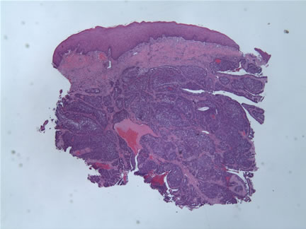
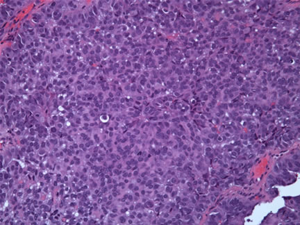
HISTOLOGICAL TYPES CHARACTERIZATION General Nonencapsulated infiltrative borders with bland regular nuclei and variable growth pattern:
Tubular
Solid
Papillary
Microcystic
Cribriform
Pseudo-adenoid cystic
Fascicular
Single file
Strand-like
- Polymorphous low-grade adenocarcinoma of the palate: report of cases.
Gonzalez-Garcia R, Rodriguez-Campo FJ, Munoz-Guerra MF, Nam-Cha SH, Sastre-Perez J, Naval-Gias L.
Department of Oral and Maxillofacial Surgery, University Hospital La Princesa, Autonoma University, c/ Diego de Leon, 62, 28006 Madrid, Spain.
Auris Nasus Larynx. 2005 Sep;32(3):275-80. Abstract quote
OBJECTIVE: Polymorphous low-grade adenocarcinoma (PLGA) is a rare tumor that mostly affects minor salivary glands. The purpose of this study is to report six new cases followed-up during a long period. We also review the literature concerning clinical, histological and immunohistochemical features, as well as the proper management.
METHODS: Malignant tumors of the salivary glands diagnosed in our department from 1990 to 1999 were reviewed. A total of 66 cases were registered. Six of these cases were diagnosed as PLGA. All cases satisfied the histopathological criteria for this entity, and at least 3 years follow-up was available.
RESULTS: In the six cases the primary location was the mucosa of the palate. Hard palate was affected in 83.3% of the cases. There were no cases of extraoral PLGA in our series. Tumors were ulcerated in a 16.6% of the cases, and exofitic in the other 86.4%. Histologically, it was observed a tumoral proliferation of round clusters of uniform cells with round-to-oval clear nuclei and small nucleoli. All the cases underwent surgical management with local excision with surgical margins, five of them with bone extirpation associated. No recurrence was observed in four cases, whereas the remaining two cases showed recurrence in the follow-up. In one of the patients, lococervical recurrence appeared 12 months after the surgery, and this patient died after a few months. The remaining patients have been followed-up for 11, 7, 4 and 3 years postoperatively, with no evidence of recurrence.
CONCLUSION: Our results for a long follow-up period support the idea for a low grade of malignancy and good prognosis of this tumor, but the appearance of recurrences many years after the surgery must induce us to be very careful and systematic with the follow-up.Polymorphous Low-Grade Adenocarcinoma A Study of 40 Cases With Long-Term Follow Up and an Evaluation of the Importance of Papillary Areas
Harry L. Evans, M.D.; Mario A. Luna, M.D.
From the Department of Pathology, The University of Texas M.D. Anderson Cancer Center, Houston, Texas, U.S.A.
Am J Surg Pathol 2000;24:1319-1328 Abstract quote
Forty cases of polymorphous low-grade adenocarcinoma with a minimum of 10 years of follow up were reviewed.
The patients included 13 men and 27 women age 22 to 71 years (median age, 54 years); 30 were white and 10 were black. The tumors were all intraoral, and the palate was the most common site (n = 24). Histologically, the neoplasms were characterized by nonencapsulated, infiltrative borders; bland, regular nuclei; and highly variable growth patterns, including tubular, solid, papillary, microcystic, cribriform (with true lumens), pseudoadenoid cystic (without true lumens), fascicular, single file, and strand-like. Papillary areas of more than focal extent were present in 17 cases, but these cases were otherwise similar to the remainder and were considered to form part of the spectrum of polymorphous low-grade adenocarcinoma. Thirteen patients had local recurrence, which was not controlled by subsequent treatment in six; six patients had cervical lymph node metastasis; three patients had distant metastasis; and five patients died of or with tumor after prolonged periods.
There was a statistically significant relationship between more than focal papillary growth and cervical lymph node metastasis, and between positive or unknown surgical margins and local recurrence (although not uncontrolled local recurrence); however, these were the only independent statistically significant correlations found between any clinical or pathologic parameter and any aspect of tumor behavior or patient survival.
VARIANTS
Polymorphous low-grade adenocarcinoma of the salivary glands with transformation to high-grade carcinoma.
Simpson RH, Pereira EM, Ribeiro AC, Abdulkadir A, Reis-Filho JS.
Department of Histopathology, Royal Devon and Exeter Hospital, Exeter, UK.
Histopathology. 2002 Sep;41(3):250-9. Abstract quote
AIMS: Polymorphous low-grade adenocarcinoma of the minor salivary glands is an infiltrative neoplasm characterized by bland-looking tumour cells arranged in diverse architectural patterns. It is considered to be of low-grade malignant potential in that nodal metastases are seen in only a minority, and distant spread is rare. Even more unusual is the transformation of polymorphous low-grade adenocarcinoma to a histologically high-grade carcinoma, i.e. dedifferentiation. In this paper, we describe the clinicopathological and immunohistochemical findings in two further examples.
METHODS AND RESULTS: Two patients presented each with a tumour of the palate. Histopathological examination showed the typical morphological, cytological and immunohistochemical features of a polymorphous low-grade adenocarcinoma. In one case there was a second component of high-grade carcinoma showing nuclear atypia, markedly increased mitotic activity and MIB1 index, as well as prominent zones of necrosis. It expressed epithelial markers and androgen receptors, and thus resembled salivary duct carcinoma. Similar tumour tissue was observed in one of the cervical nodal metastases, which was biopsied at the same time as the palate. In the second patient, a high-grade component was discovered when the tumour recurred in the palate 13 years after the initial biopsy. Whilst morphologically similar to that in first case, there were significant immunohistochemical differences such as retention of some of the polymorphous low-grade adenocarcinoma profile and absence of androgen receptor expression.
CONCLUSIONS: Polymorphous low-grade adenocarcinoma was first described relatively recently, and as experience with it continues to accumulate, it is becoming clear that late recurrences and metastases, whilst still infrequent, may not be quite as rare as previously thought. Reports of histological transformation are even scarcer, and most occurred at least 13 years after the polymorphous low-grade adenocarcinoma was initially recognized. It is a real possibility that this phenomenon, like clinical progression, may also be encountered more often as time passes. Therefore, we believe that, whilst polymorphous low-grade adenocarcinoma is certainly far less aggressive than, for example, adenoid cystic carcinoma, it nevertheless remains a true malignancy with a potential to prove fatal in a minority of patients.
Histologic transformation of polymorphous low-grade adenocarcinoma of salivary gland.
Pelkey TJ, Mills SE.
Department of Pathology, University of Virginia Medical Center, Charlottesville 22908, USA.
Am J Clin Pathol 1999 Jun;111(6):785-91 Abstract quote
Polymorphous low-grade adenocarcinoma of salivary gland origin (PLGA) was initially described in 1984 and has since become an established clinicopathologic entity. Owing to the indolent nature of PLGA and its relatively recent description, the full clinicopathologic spectrum of this entity has not been elucidated fully. Transformation to a histologically different-appearing lesion or progression to a higher histologic grade has not been reported.
We describe 2 PLGAs arising in the palate and associated with multiple locoregional recurrences that were treated with excision and radiation therapy. This was followed by histologic transformation to a higher grade neoplasm after 17 and 26 years, respectively. The histologic appearance after transformation was characterized by a predominantly solid and cystic growth pattern, nuclear atypia with prominent nucleoli, and foci of necrosis. High-grade transformation of PLGA may occur after a protracted clinical course with multiple recurrences of typical PLGA.
The possible role of radiation therapy as an initiator of this transformation merits further study. Tumors with these histologic features should not be included under the rubric of typical PLGA. Segregation of these neoplasms will allow further study of their biologic potential, particularly with regard to possible increased rates of local recurrence and metastasis.
SPECIAL STAINS/
IMMUNOPEROXIDASEGENERAL
- Beta-6 Integrin, tenascin-C, and MMP-1 expression in salivary gland neoplasms.
Westernoff TH, Jordan RC, Regezi JA, Ramos DM, Schmidt BL.
Department of Oral and Maxillofacial Surgery, 521 Parnassus Avenue, C-522, University of California, San Francisco, CA 94143-0440, United States.
Oral Oncol. 2005 Feb;41(2):170-4. Abstract quote
Beta-6 Integrin, tenascin-C, and MMP-1 (matrix metalloproteinase-1) are invasion-related proteins that are frequently overexpressed in many human malignancies.
The objective of this study was to determine whether there is overexpression of these molecules in three types of salivary neoplasms showing markedly different behavior.
A total of 55 formalin-fixed, paraffin-embedded archived specimens comprising 19 adenoid cystic carcinomas (ACC), 18 polymorphous low-grade adenocarcinomas (PLGA) and 18 pleomorphic adenomas (PA) were utilized in this study. A standard immunohistochemical technique was used to determine the expression levels of beta-6 integrin, tenascin-C, and matrix metalloproteinase-1 (MMP-1) proteins. Sections were assessed semiquantitatively, and tumors were divided into two groups, low-expressors (0-1+) and high-expressors (2-3+) for statistical analysis. Staining was graded as 0 (<1% positive tumor cells), 1+ (<25% positive tumor cells), 2+ (25-50% positive tumor cells), and 3+ (>50% positive cells). The results showed that the malignant tumors were higher expressors of beta-6 than the benign tumors. ACCs showed significantly higher expression of beta-6 than PAs (p=0.04). No significant difference was observed between ACCs and PLGAs. beta-6 expression was rarely seen in normal salivary gland epithelium and was occasionally present in mucosa overlying the tumors.
PAs were high-expressors of tenascin-C with a significant difference relative to ACCs (p=0.03). A majority of tumors in all three tumor types showed high expression of MMP1 with expression significantly greater in the PAs compared to ACCs (p=0.008).
We conclude that ACCs and PLGAs express beta-6, tenascin-C, and MMP-1, but that their expression patterns are not significantly different. beta-6 appears to be more closely associated with the malignant tumors, and MMP-1 more closely associated with the benign tumors. We believe that beta-6, tenascin-C, and MMP-1 proteins are part of the molecular repertoire used by salivary tumors for malignant invasion and benign tumor expansion.CD117 (c-kit)
- C-kit expression in the salivary gland neoplasms adenoid cystic carcinoma, polymorphous low-grade adenocarcinoma, and monomorphic adenoma.
Edwards PC, Bhuiya T, Kelsch RD.
Department of Dental Medicine, Long Island Jewish Medical Center, 270-05 76th Avenue, New Hyde Park, NY 11040, USA.
Oral Surg Oral Med Oral Pathol Oral Radiol Endod. 2003 May;95(5):586-93. Abstract quote
OBJECTIVE: Differentiating between adenoid cystic carcinomas (ACCs), polymorphous low-grade adenocarcinomas (PLGAs), and the monomorphic adenomas (including canalicular adenomas, trabecular adenomas, and basal cell adenomas) can present a diagnostic challenge, especially when examining tissue obtained from small incisional or fragmented biopsies. Recent studies have revealed that overexpression of the tyrosine kinase receptor protein c-kit occurs in a narrow subset of malignant neoplasms, including gastrointestinal stromal tumors, myeloid leukemias, seminomas, and ACCs. C-kit reportedly is not expressed in PLGAs. We compared the expression of the c-kit antigen in the malignant salivary gland neoplasms ACC and PLGA with its expression in salivary gland monomorphic adenoma (including canalicular adenoma and basal cell adenoma).
STUDY DESIGN: Formalin-fixed paraffin-embedded sections of 49 salivary gland neoplasms (17 monomorphic adenomas, 17 PLGAs, and 15 ACCs) accessioned between 1989 and 2002 were retrieved from the files of the Department of Pathology, Long Island Jewish Medical Center, and were stained with an anti-c-kit polyclonal antibody.
RESULTS: C-kit reactivity was uniformly positive in the cytoplasm of luminal neoplastic cells in ACCs (15/15, 100%). Positive reactivity was also identified in the majority of PLGAs (16/17, 94%), with at least 25% of the tumor cells being positive. Similar reactivity was seen in monomorphic adenomas (16/17, 94%).
CONCLUSIONS: In contrast to previous reports, we find that c-kit expression was not restricted to ACC but was expressed in all 3 tumor types evaluated (ACC, PLGA, and monomorphic adenoma). Therefore, c-kit does not appear to be a useful marker for distinguishing between either ACC and PLGA in equivocal cases, or in benign and malignant salivary gland neoplasms.
- C-kit expression distinguishes salivary gland adenoid cystic carcinoma from polymorphous low-grade adenocarcinoma.
Penner CR, Folpe AL, Budnick SD.
Department of Pathology and Laboratory Medicine, Emory University, Atlanta, Georgia 30322, USA.
Mod Pathol. 2002 Jul;15(7):687-91. Abstract quote
Adenoid cystic carcinoma (ACC) is characterized by persistent, relentless growth and a high rate of eventual metastasis. In contrast, polymorphous low-grade adenocarcinoma (PLGA) has a much lower risk of recurrence and rarely metastasizes. The histologic patterns of these two neoplasms can be similar. Expression of c-kit, a transmembrane receptor tyrosine kinase, has recently been reported to be expressed in ACC but not PLGA. Expression of galectin-3, a nonintegrin beta-galactosidase-binding lectin, has been reported to be significant in PLGA and decreased in ACC.
Formalin-fixed paraffin-embedded tissue from 9 ACC and 14 PLGA were immunostained for c-kit and galectin-3. Cases were scored as 1+ (5-25% positive), 2+ (26-50% positive), or 3+ (>50% positive). C-kit was expressed by 100% of ACC (3+: 7 cases; 2+: 1 case; 1+: 1 case) and by 57% of PLGA (2+: 2 cases; 1+: 6 cases). In all but one ACC, c-kit expression was confined to the inner cell layer. C-kit expression was also noted in the intercalated duct epithelium of the salivary glands and the acinar cells of the lacrimal gland. Galectin-3 was expressed in 8 of 9 cases of ACC and 14 of 14 cases of PLGA.
The results of this, the first study to compare c-kit and galectin-3 expression in ACC and PLGA, suggest that c-kit expression characterizes ACC, but not PLGA. Galectin-3 immunohistochemistry does not have a role in the differentiation of ACC and PLGA. C-kit immunostaining may be a valuable adjunctive tool for this differential diagnosis, particularly in the setting of a limited biopsy.
Our finding of different patterns of c-kit expression in tubular and solid variants of ACC supports the concept of solid variant ACC as a high-grade tumor, with progression toward an entirely "inner cell" phenotype.CYTOKERATINS
- Immunohistochemical expression of cytokeratins 7 and 20 in malignant salivary gland tumors.
Nikitakis NG, Tosios KI, Papanikolaou VS, Rivera H, Papanicolaou SI, Ioffe OB.
Department of Diagnostic Sciences and Pathology, University of Maryland, Baltimore, MD 21201, USA.
Mod Pathol. 2004 Apr;17(4):407-15. Abstract quote
On the basis of the heterogeneity of cytokeratins 7 and 20 expression in malignant epithelial tumors, the cytokeratin 7/20 immunophenotype has served as a useful diagnostic tool for discrimination of primary and/or metastatic carcinomas of unknown origin. However, the expression pattern of these cytokeratins in malignant salivary gland tumors has not been thoroughly studied.
Our study material was composed of 84 malignant tumors of primary major or minor salivary gland origin. Nine histologic types of carcinoma were represented, including mucoepidermoid (26 cases), adenoid cystic (25), polymorphous low grade (11), salivary duct (8), acinic cell (4), ex mixed tumor (3), not otherwise specified (3), clear cell (2), and basal cell (2). In all, 13 cases of primary skin or mucosal squamous cell carcinoma with secondary salivary gland involvement were also examined. Immunoreactivity for cytokeratin 7 was evident in all malignant salivary gland tumors; the staining pattern was diffuse and strong in 62 cases, and focal and strong in 22 cases. In contrast, 78 cases were negative for cytokeratin 20, whereas only six cases (two mucoepidermoid, one adenoid cystic, and three salivary duct) displayed focal weak positivity.
Overall, 92.9% of malignant salivary gland tumors were characterized by a cytokeratin 7 positive/20 negative immunoprofile, the remaining 7.1% of cases being positive for both cytokeratins. The latter phenotype was more common in salivary duct carcinomas (P< or =0.05). On the other hand, most squamous cell carcinomas (69%) were negative for both cytokeratins, while the remaining cases (31%) were negative for cytokeratin 20 and focally weakly positive for cytokeratin 7.
We suggest that assessment of cytokeratin 7/20 immunoprofile may facilitate the differential diagnosis of (a) primary malignant salivary gland tumors from metastatic tumors, (b) metastatic salivary gland tumors, (c) primary salivary gland tumors, especially mucoepidermoid carcinomas, from squamous cell carcinomas, and (d) salivary duct carcinomas from other malignant salivary gland tumors.p63
- Assessment of p63 expression in the salivary gland neoplasms adenoid cystic carcinoma, polymorphous low-grade adenocarcinoma, and basal cell and canalicular adenomas.
Edwards PC, Bhuiya T, Kelsch RD.
Department of Dental Medicine, Division of Oral Pathology, Long Island Jewish Medical Center, New Hyde Park, NY 11040, USA.
Oral Surg Oral Med Oral Pathol Oral Radiol Endod. 2004 May;97(5):613-9. Abstract quote
PURPOSE: The purpose of this study was to determine the extent of p63 immunoreactivity in the malignant salivary gland neoplasms adenoid cystic carcinoma (ACC) and polymorphous low-grade adenocarcinoma (PLGA) and to compare this to the expression of this marker in the benign salivary gland tumors canalicular adenoma and basal cell adenoma. Few studies on the expression of p63 in head and neck salivary gland tumors have been published to date. P63, a selective immunohistochemical marker of basal/stem cells of stratified epithelium and of myoepithelial cells, is a p53 homologue that plays an essential role in both morphogenesis of epidermis and limb development. P63 immunoreactivity has been demonstrated in squamous cell and urothelial carcinomas. It is generally absent in most nonsquamous cell carcinomas.Study design Formalin-fixed paraffin-embedded sections from 49 salivary gland neoplasms, representing 6 canalicular adenomas, 11 basal cell adenomas, 17 PLGA and 15 ACC accessioned from 1989 to 2002 by the Department of Pathology, Long Island Jewish Medical Center, New Hyde Park, NY, were stained with an anti-p63 monoclonal antibody.
RESULTS: Nuclear p63 reactivity was uniformly positive in PLGA (17/17, 100%). Positive reactivity was also identified in the majority of cases of ACC (13/15, 87%), primarily in the nonluminal myoepithelial-like cells surrounding luminal cells. Canalicular adenoma did not exhibit any p63 immunoreactivity. All basal cell adenomas of parotid origin stained strongly for p63, with staining localized to the peripheral tumor cells situated adjacent to the connective tissue stroma. None of the basal cell adenomas originating in the upper lip stained with p63. In native adjacent salivary gland tissue, p63 reactivity was identified focally in the nuclei of myoepithelial and basal duct cells.
CONCLUSIONS: P63 is strongly expressed in basal cell adenoma of parotid origin, and in ACC and PLGA. Canalicular adenoma did not demonstrate p63 staining, consistent with this tumor's putative luminal ductal cell differentiation. Our results suggest that the neoplastic cells in PLGA may represent either a population of p63-positive epithelial stem/reserve cells similar to the basal cells of stratified epithelium, or modified myoepithelial cells. Given the staining pattern of the tumors examined, p63 does not appear to be an ideal marker for distinguishing between ACC, PLGA, and basal cell adenoma.
DIFFERENTIAL DIAGNOSIS KEY DIFFERENTIATING FEATURES ADENOID CYSTIC CARCINOMA
- Selective immunohistochemical comparison of polymorphous low-grade adenocarcinoma and adenoid cystic carcinoma.
Beltran D, Faquin WC, Gallagher G, August M.
Department of Oral and Maxillofacial Surgery, Massachusetts General Hospital, Harvard School of Dental Medicine, Boston, MA 02114, USA.
J Oral Maxillofac Surg. 2006 Mar;64(3):415-23. Abstract quote
PURPOSE: Polymorphous low-grade adenocarcinoma (PLGA) is a salivary gland malignancy characterized by indolent growth and a low rate of metastasis. PLGA shares histological features with adenoid cystic carcinoma (ACC), including infiltrating solid and cribriform patterns, presence of cystic spaces, and neurotropism. The degree of polymorphism of PLGA presents diagnostic challenges, particularly in small biopsy specimens. Immunohistochemical reactions to differentiate PLGA from the more aggressive ACC would be extremely valuable but controversy exists in the current literature regarding their utility. This study examines the potential use of c-kit, Ki-67, smooth muscle actin (SMA), and muscle-specific actin (MSA) as ancillary markers for distinguishing PLGA from ACC.
PATIENTS AND METHODS: Medical records of 20 cases of PLGA treated at the Massachusetts General Hospital were reviewed. Patient demographic data and tumor-specific information were elicited. Formalin-fixed paraffin-embedded sections from 10 of these cases and 12 comparison cases of ACC were accessed. The histologic diagnoses were confirmed and immunohistochemical staining using antibodies to c-kit, Ki-67, SMA, and MSA was employed to determine differences in staining.
RESULTS: PLGA showed a significantly weaker immunohistochemical expression of c-kit compared with ACC (P = < .001). Ki-67, correlating with proliferative behavior, was more weakly expressed in PLGA (P = .091). The exuberant myoepithelial component of ACC resulted in stronger staining with SMA and MSA (P = .047; P = .065, respectively).
CONCLUSIONS: Statistically significant immunohistochemical staining patterns using c-kit and SMA in this study support their potential use as markers to differentiate PLGA from ACC in cases where the diagnosis can be challenging.
- Adenoid cystic carcinoma and polymorphous low-grade adenocarcinoma of minor salivary glands: a comparative immunohistochemical study using the epithelial membrane and carcinoembryonic antibodies.
Epivatianos A, Iordanides S, Zaraboukas T, Antoniades D.
Department of Oral Medicine and Oral Pathology, Universit of Thessaloniki, Thessaloniki, Greece.
Oral Dis. 2005 May;11(3):175-80. Abstract quote
OBJECTIVE: The purpose of this study was to investigate immunohistochemically the expression of epithelial membrane antigen (EMA) and carcinoembryonic antigen (CEA) in adenoid cystic carcinoma (AdCC) and polymorphous low-grade adenocarcinoma (PLGA) in an attempt to assess the ability of these markers to distinguish AdCC from PLGA when the histological features on routine hematoxylin and eosin are equivocal.
MATERIALS AND METHODS: Fourteen specimens of AdCC, 10 PLGA, and five normal minor salivary glands fixed in 10% formalin and embedded in paraffin, were retrieved from the files of our department and were retrospectively studied with the streptavidin-biotin complex method using the epithelial membrane and carcinoembryonic antibodies.
RESULTS: The immunoreactivities and the expression patterns of EMA and CEA in AdCC and PLGA were similar.
CONCLUSIONS: The results of this study suggest that the immunostaining of AdCC and PLGA with EMA and CEA could not offer an adjunctive aid in differential diagnosis between these two tumors.The cribriform features of adenoid cystic carcinoma and polymorphous low-grade adenocarcinoma: Cytokeratin and integrin expression
Vera C. Ara�jo, DDS, PhD
Silvia V.L. Loducca, DDS, PhD
Suzana O.M. Sousa, DDS, PhD
David M. Williams, DDS, PhD
Ney S. Ara�jo, DDS, PhDAnn Diagn Pathol 2001;5:330-334 Abstract quote
Cribriform areas are common features of both adenoid cystic carcinoma and polymorphous low-grade adenocarcinoma. Both are malignant salivary gland tumors that share similar histologic patterns, but with marked distinct clinical behavior.
This study was undertaken to improve the accuracy of the histopathology diagnostic process, using an immunohistochemical panel to differentiate adenoid cystic carcinoma from polymorphous low-grade adenocarcinoma, with special concern to the common cribriform areas shared by these tumors. Three-�m serial sections of these tumors were submitted to the streptavidin-biotin peroxidase immunotechnique against the monoclonal antibodies anticytokeratins 7, 8, 14 and 19, and anti-integrins 1, 3, and 4. In the neoplastic lobules of adenoid cystic carcinoma cribriform type, the spaces were mainly surrounded by cells negative for the cytokeratins and integrins studied. In the solid type of adenoid cystic carcinoma, the microcystic areas were caused by spaces lined by neoplastic luminal cells positive for cytokeratins and presenting integrins concentrated in the apical pole of these cells. The cribriform areas of polymorphous low-grade adenocarcinoma were composed of cords of luminal cells, positive for cytokeratins and showing integrins disposed in a bipolar pattern.
We concluded that cribriform areas of adenoid cystic carcinoma and polymorphous low-grade adenocarcinoma are histologically similar, although not identical. Indeed, their cellular composition is distinct and can be distinguishable from one another by the proteins of the cytoskeleton, by the integrins, or both.
C-kit Expression Distinguishes Salivary Gland Adenoid Cystic Carcinoma from Polymorphous Low-Grade Adenocarcinoma.Penner CR, Folpe AL, Budnick SD.
Department of Pathology and Laboratory Medicine, Emory University, Atlanta, Georgia.
Mod Pathol 2002 Jul;15(7):687-91 Abstract quote Adenoid cystic carcinoma (ACC) is characterized by persistent, relentless growth and a high rate of eventual metastasis. In contrast, polymorphous low-grade adenocarcinoma (PLGA) has a much lower risk of recurrence and rarely metastasizes. The histologic patterns of these two neoplasms can be similar. Expression of c-kit, a transmembrane receptor tyrosine kinase, has recently been reported to be expressed in ACC but not PLGA. Expression of galectin-3, a nonintegrin beta-galactosidase-binding lectin, has been reported to be significant in PLGA and decreased in ACC.
Formalin-fixed paraffin-embedded tissue from 9 ACC and 14 PLGA were immunostained for c-kit and galectin-3. Cases were scored as 1+ (5-25% positive), 2+ (26-50% positive), or 3+ (>50% positive). C-kit was expressed by 100% of ACC (3+: 7 cases; 2+: 1 case; 1+: 1 case) and by 57% of PLGA (2+: 2 cases; 1+: 6 cases). In all but one ACC, c-kit expression was confined to the inner cell layer. C-kit expression was also noted in the intercalated duct epithelium of the salivary glands and the acinar cells of the lacrimal gland. Galectin-3 was expressed in 8 of 9 cases of ACC and 14 of 14 cases of PLGA.The results of this, the first study to compare c-kit and galectin-3 expression in ACC and PLGA, suggest that c-kit expression characterizes ACC, but not PLGA. Galectin-3 immunohistochemistry does not have a role in the differentiation of ACC and PLGA. C-kit immunostaining may be a valuable adjunctive tool for this differential diagnosis, particularly in the setting of a limited biopsy.
Our finding of different patterns of c-kit expression in tubular and solid variants of ACC supports the concept of solid variant ACC as a high-grade tumor, with progression toward an entirely "inner cell" phenotype.
CANALICULAR ADENOMA
Comparative immunoprofile of polymorphous low-grade adenocarcinoma and canalicular adenoma.
Furuse C, Tucci R, Machado De Sousa SO, Rodarte Carvalho Y, Cavalcanti De Araujo V.
Ann Diagn Pathol. 2003 Oct;7(5):278-80. Abstract quote Immunohistochemistry is an important tool when dealing with salivary gland neoplasms. Canalicular adenoma and polymorphous low-grade adenocarcinoma may share some histologic characteristics that can cause difficulties in their separation.
In the present study, cases of polymorphous low-grade adenocarcinoma and canalicular adenoma were submitted to a panel of antibodies to evaluate the differences in their immunoprofiles. The results obtained showed that, while vimentin is only expressed by polymorphous low-grade adenocarcinoma, CK7 and CK8 are present in both neoplasms.
Therefore, vimentin is the best marker to differentiate between these tumors.
METASTASTIC LOBULAR CARCINOMA OF THE BREAST Lobular carcinoma of the breast metastatic to the oral cavity mimicking polymorphous low-grade adenocarcinoma of the minor salivary glands.
Nicol KK, Iskandar SS.
Department of Pathology, Wake Forest University School of Medicine, Winston-Salem, NC 27157-1072, USA.
Arch Pathol Lab Med 2000 Jan;124(1):157-9 Abstract quote
The oral cavity is a rare site of metastatic lesions; however, metastatic breast carcinoma must be included in the differential diagnosis of tumors of that site in women.
We describe a 54-year-old woman who presented with a lesion of the floor of the mouth that histologically resembled polymorphous low-grade adenocarcinoma of the minor salivary glands, which was eventually established to represent metastatic lobular breast carcinoma.
The final diagnosis was based on comparison with a primary tumor resected 13 years earlier and immunohistochemical reactivity with antibodies to steroid receptors. Relevant aspects of lobular breast carcinoma, polymorphous low-grade adenocarcinoma, and metastatic oral cavity lesions are discussed.
PROGNOSIS AND TREATMENT CHARACTERIZATION PROGNOSIS Am J Surg Pathol 2000;24:1319-1328
17/40 cases had papillary foci13/17 had local recurrence which was not controlled by subsequent treatment in 6 cases
3/17 had distant metastasis
5/17 died of or with tumor after prolonged periods
Statistically significant association between more than focal papillary growth and cervical lymph node metastasis
Positive margins have increased risk of local recurrence Survival See above RECURRENCE See above
Recurrent polymorphous low-grade adenocarcinoma manifesting as a sinonasal mass: a case report.
Charous DD, Cunnane MF, Rosen MR, Keane WM.
Department of Otolaryngology-Head and Neck Surgery, Thomas Jefferson University, 925 Chestnut St., Philadelphia, PA 19107, USA.
Ear Nose Throat J. 2005 Jun;84(6):354, 356-7. Abstract quote
Polymorphous low-grade adenocarcinoma (PLGA) is a malignant neoplasm that tends to follow a benign clinical course. Recurrences are uncommon.
We report a case of recurrent PLGA of the paranasal sinuses that manifested as a large mass that filled the entire nasal cavity and left maxillary sinus.
To our knowledge, this is the first reported case of a recurrent PLGA of the paranasal sinuses.
Metastasis Cervical lymph nodes
Ilium
Lungs
SkinCell proliferation in salivary gland adenocarcinomas with myoepithelial participation. A study of 78 cases.
Fonseca I, Felix A, Soares J.
Departamento de Patologia Morfologica & Centro de Investigacao de Patobiologia Molecular (CIPM), Instituto Portugues de Oncologia de Francisco Gentil, Lisboa, Portugal.
Virchows Arch 1997 Mar;430(3):227-32 Abstract quote
We used three markers of cell proliferation mitotic counts, mitotic index and expression of proliferating cell nuclear antigen--to assess the proliferative activity of a series of 78 low-grade salivary adenocarcinomas with myoepithelial participation classified according to: their histological type, the predominant architectural type, and the predominant cytological type. The series included adenoid cystic carcinomas (40), epithelial-myoepithelial carcinomas (19), polymorphous low-grade adenocarcinomas (12) and basal cell adenocarcinomas (7).
The proliferation indicators were found to be similar in the first three groups, being significantly lower than in the last. Tumours formed by basal cells had statistically significant higher mitotic indexes than those predominantly composed of clear cells of myoepithelial type and ductal cells. Tubular tumours, irrespective of the histological classification of the neoplasm, had proliferation indexes similar to those found in cribriform neoplasms. Solid tumours, whether formed by ductal or clear myoepithelial-type cells, had higher indexes than the neoplasms with differentiated (cribriform and tubular) patterns. The highest mean values for every proliferation indicator used were found in tumours with solid organization that were predominantly formed by basal cells. These results agree with the hypothesis that cell proliferation is inversely related to neoplastic differentiation.
The identification of the prevalent cell phenotype and architecture may extend our knowledge from adenoid cystic carcinoma, whose solid variant carries a worse prognosis, and supports that the usual classification of this group of salivary adenocarcinomas would benefit to be complemented with information on tumour architecture and cellular composition.
TREATMENT Surgery often followed by radiation
Surgery varied from local excision to partial maxillectomy and mandibulectomyCervical lymph node dissection is often combined
Mod Pathol 1990;3:76-82.
Cancer 1999;86:207-219.
Am J Surg Pathol 2000;24:1319-1328.
Henry JB. Clinical Diagnosis and Management by Laboratory Methods. Twentieth Edition. WB Saunders. 2001.
Rosai J. Ackerman's Surgical Pathology. Ninth Edition. Mosby 2004.
Sternberg S. Diagnostic Surgical Pathology. Fourth Edition. Lipincott Williams and Wilkins 2004.
Robbins Pathologic Basis of Disease. Seventh Edition. WB Saunders 2005.
DeMay RM. The Art and Science of Cytopathology. Volume 1 and 2. ASCP Press. 1996.
Weedon D. Weedon's Skin Pathology Second Edition. Churchill Livingstone. 2002
Fitzpatrick's Dermatology in General Medicine. 6th Edition. McGraw-Hill. 2003.
Weiss SW and Goldblum JR. Enzinger and Weiss's Soft Tissue Tumors. Fourth Edition. Mosby 2001.
Basic Principles of Disease
Learn the basic disease classifications of cancers, infections, and inflammation
Commonly Used Terms
This is a glossary of terms often found in a pathology report.Diagnostic Process
Learn how a pathologist makes a diagnosis using a microscopeSurgical Pathology Report
Examine an actual biopsy report to understand what each section meansSpecial Stains
Understand the tools the pathologist utilizes to aid in the diagnosisHow Accurate is My Report?
Pathologists actively oversee every area of the laboratory to ensure your report is accurateGot Path?
Recent teaching cases and lectures presented in conferences
Pathologists Who Make A Difference
Search for a Physician Specialist
Microscopic Photo
Microscopic Photo
Last Updated April 13, 2006
Send
mail to The Doctor's Doctor with
questions or comments about this web site.
Read the Medical Disclaimer.
Copyright © The Doctor's Doctor